group of peopleskeletonanatomydissectionmedical illustrationcavitiesthoracicabdominaldiagramschartsclassroomlaboratoryhistorical photographvintage imageblack and whitemensuitsformal wearmedical studentsprofessorinstructorcadaverhuman bodyanatomy lessonmedical educationscientific studyresearchlearningknowledgeexpertisehistorical contextmedical historyscientific advancementhuman anatomybiological studymedical practicehealthcaremedical professionscientific inquiryhuman bodyanatomydissectionmedical illustrationdiagramschartsclassroomlaboratoryhistorical photographvintage imageblack and whitemensuitsformal wearmedical studentsprofessorinstructorcadaverhuman bodyanatomy lessonmedical educationscientific studyresearchlearningknowledgeexpertisehistorical contextmedical historyscientific advancementhuman anatomybiological studymedical practicehealthcaremedical professionscientific inquiryanatomydissectionmedicaleducationhistoricalvintageblack and whiteclassroomlaboratorymensuitsmedical studentsprofessorinstructorcadaverthoracicabdominalcavitiesdiagramscharts
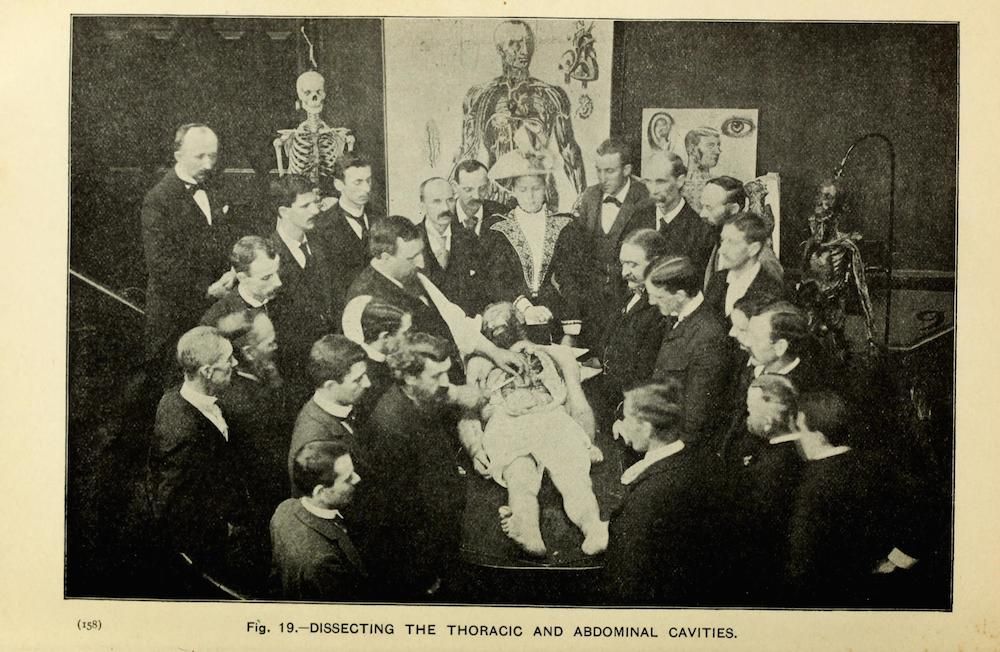
This is a vintage black and white photograph depicting a medical anatomy lesson. A large group of men, all dressed in suits and formal wear, are gathered around a cadaver that is being dissected. The cadaver is lying on a table, and several individuals are actively engaged in the dissection process. In the background, there are anatomical charts and diagrams displayed on the walls, illustrating the thoracic and abdominal cavities. The scene appears to be taking place in a classroom or laboratory setting, likely a medical school. The photograph captures a moment of intense scientific study and learning, showcasing the historical practice of anatomical dissection in medical education. The image is labeled 'Fig. 19. - DISSECTING THE THORACIC AND ABDOMINAL CAVITIES.'
License: CC0