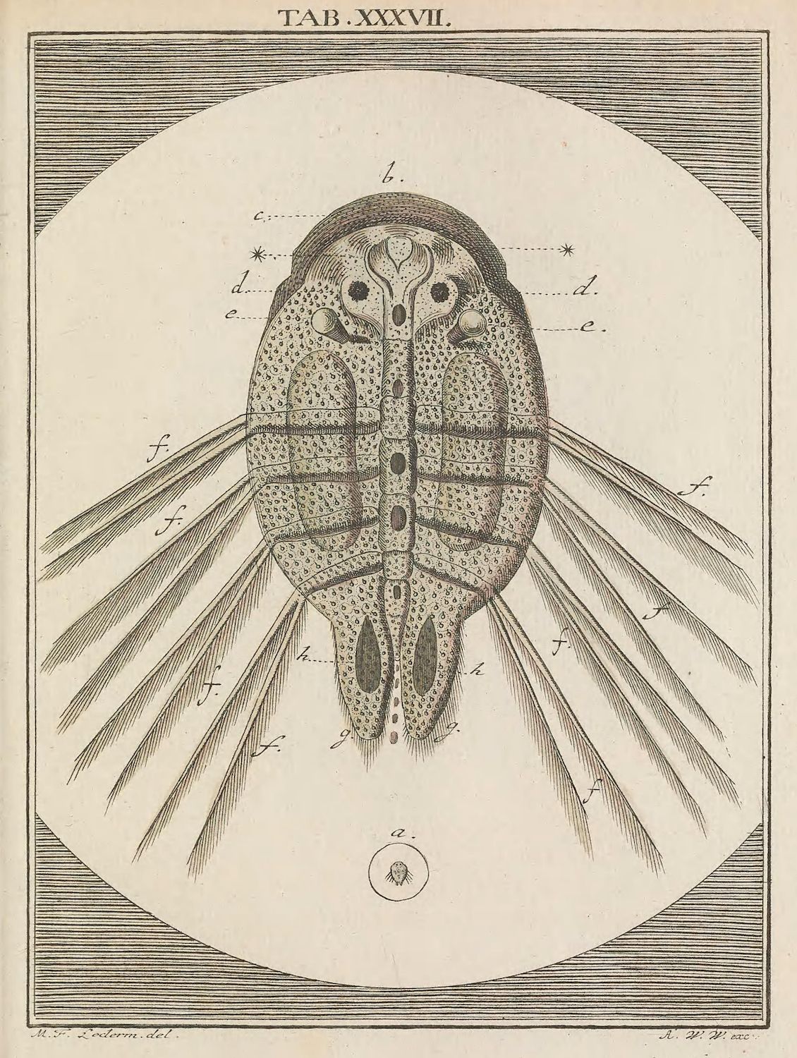
This is a detailed scientific illustration of a water flea, likely from a historical scientific text. The image is a black and white engraving, with a circular border. The central focus is a large, oval-shaped water flea, depicted in a dorsal view. The body is filled with intricate stippling to represent texture and internal structures. Several labels with letters (a, c, d, e, f, g, h, i) point to different parts of the creature's anatomy. Key features include: * **Body:** The oval body is the most prominent feature, with detailed internal organs visible through the translucent shell. * **Antennae:** Two antennae (labeled 'c') extend from the head. * **Legs/Appendages:** Numerous thin, leg-like appendages (labeled 'f') radiate outwards from the lower part of the body, suggesting the creature's method of locomotion or feeding. * **Internal Structures:** The illustration shows a complex internal structure, including what appears to be a digestive tract and other organs. * **Labels:** The letters 'a', 'c', 'd', 'e', 'f', 'g', 'h', and 'i' are used to label specific parts of the water flea's anatomy. * **Small Circle:** A small circle with a dot in the center is located at the bottom of the image, labeled 'a'. Above the illustration, the text “TAB. XXVII.” is visible, likely indicating the plate number in a larger publication. The bottom of the image includes the names of the artist and engraver, “M.F. Seiden del.” and “A.W.Y. exc.” respectively. The overall style is characteristic of 18th or 19th-century scientific illustrations, emphasizing detail and accuracy.