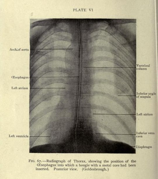
The image is a vintage radiograph of a thorax, labeled as 'Plate VI' and 'Fig. 67'. It's a posterior view, showing the skeletal structure and internal organs. The radiograph is grayscale, with varying shades of gray indicating different densities. A prominent, bright, linear structure runs down the center, representing a bougie with a metal core inserted into the esophagus. The ribs are visible as curved, darker lines radiating from the spine. The heart and great vessels are discernible, with labels pointing to the left atrium, left ventricle, and arch of aorta. The spine is visible as a vertical, darker line. Labels also indicate the inferior angle of scapula, inferior vena cava, and diaphragm. The image has a slightly aged appearance, with some imperfections and discoloration. The overall impression is that of a historical medical illustration.