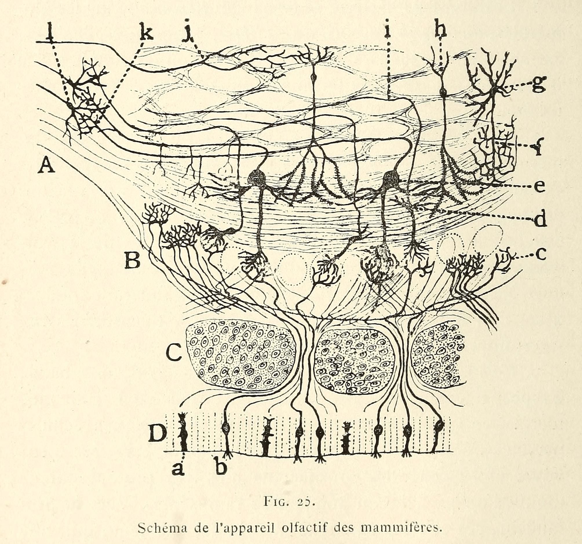
This is a vintage scientific diagram illustrating the olfactory system of mammals. It's a detailed cross-section, likely intended for anatomical study. The diagram is labeled with letters and lines to indicate different structures. Here's a breakdown of the key elements: * **Layers:** The diagram shows several layers, labeled A, B, C, and D, representing different parts of the olfactory system. * **Olfactory Receptor Neurons:** In layer D, there are numerous small, elongated cells with cilia (hair-like structures) extending into the nasal cavity. These are the olfactory receptor neurons. * **Olfactory Bulb:** Layer C shows the olfactory bulb, a bulbous structure with a layered appearance. Within the bulb, there are clusters of cells (glomeruli) where olfactory information is processed. * **Mitral Cells:** Layer B depicts mitral cells, larger neurons with extensive branching dendrites that receive input from the olfactory receptor neurons. * **Olfactory Tract:** Layer A shows the olfactory tract, a bundle of nerve fibers that carries olfactory information from the olfactory bulb to the brain. * **Labels:** The diagram is heavily labeled with letters (a, b, c, d, e, f, g, h, i, j, k, l) and lines pointing to specific structures. These labels likely correspond to a key or legend explaining the different parts of the olfactory system. The style of the diagram is typical of vintage scientific illustrations, with detailed line work and a monochromatic color scheme. The overall impression is one of complexity and precision, reflecting the intricate nature of the olfactory system.