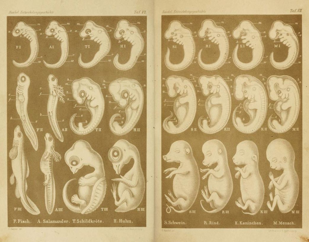
This is a vintage scientific illustration, likely from a biology or anatomy textbook. It's a comparative study of embryonic development across different species. The image is divided into two main sections, each containing a series of diagrams showing the stages of embryonic development. **Left Section (Tab. VI):** This section focuses on fish, salamander, frog, and chicken embryos. The diagrams are arranged in rows, each row representing a different stage of development (I, II, III). The embryos are depicted as translucent, with internal structures labeled with letters (a, b, c, d, etc.). The stages progress from early, simple forms to more complex, recognizable shapes. **Right Section (Tab. VII):** This section shows the embryonic development of pig, cow, rabbit, and human. Similar to the left section, the diagrams are arranged in rows (I, II, III) representing different stages. The embryos are also translucent, with internal structures labeled. The stages show a progression from early forms to more developed shapes. The overall style is detailed and precise, typical of scientific illustrations from the 19th or early 20th century. The diagrams are sepia-toned, giving the image a vintage appearance. The text at the bottom identifies the species depicted in each section: F. Fisch (fish), A. Salamander, T. Schildkröte (turtle), H. Huhn (chicken), S. Schwein (pig), R. Rind (cow), K. Kaninchen (rabbit), and M. Mensch (human).