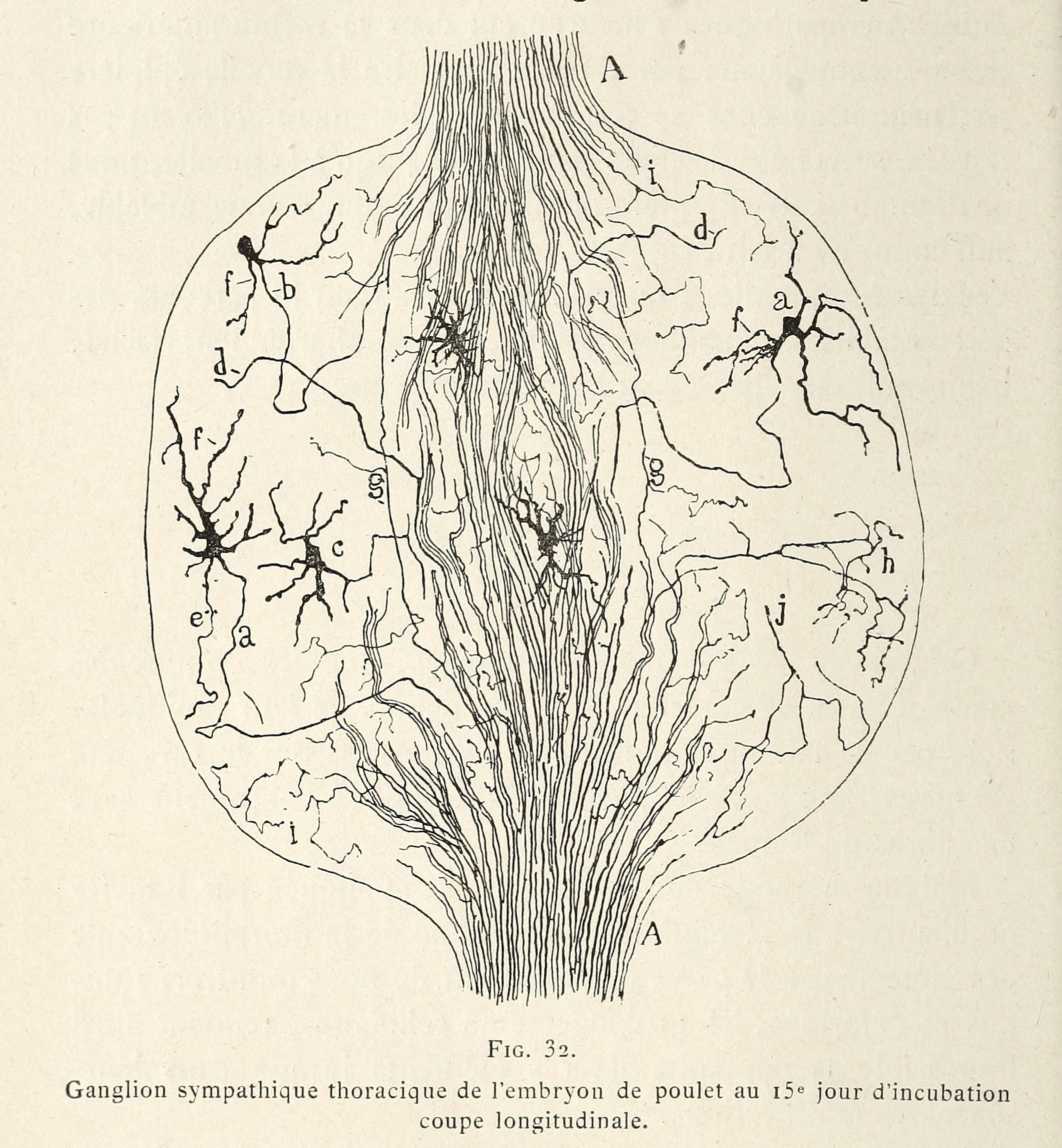
This is a detailed biological illustration, specifically a diagram of the thoracic sympathetic ganglion of a chicken embryo at 15 days of incubation, shown in longitudinal section. The diagram is rendered in black and white with fine lines and stippling. The central feature is an oval-shaped ganglion, densely packed with cells and fibers. Numerous nerve fibers radiate outwards from the ganglion, branching and interconnecting. The cells within the ganglion are depicted as small, irregular shapes. Several labels (a, b, c, d, e, f, g, h, i, j, k, l) point to specific structures within the ganglion and its surrounding fibers. The illustration is highly detailed, showing the complex network of nerve cells and fibers within the ganglion. The text below the image reads 'Ganglion sympathique thoracique de l'embryon du poulet au 15e jour d'incubation. coupe longitudinale.'