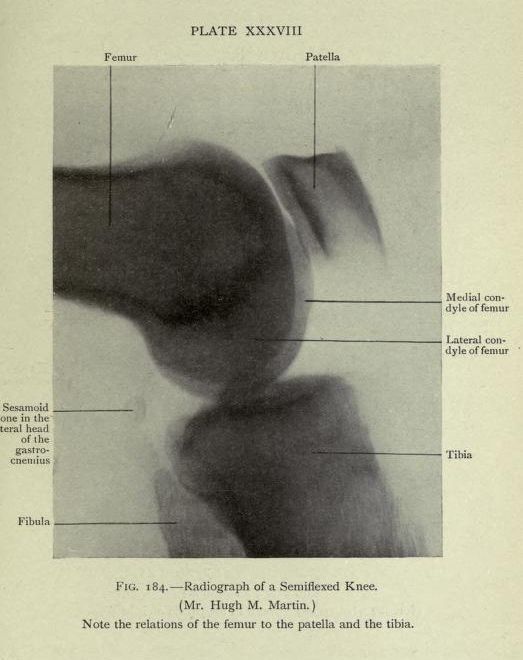
This is a vintage radiographic image of a semiflexed knee, labeled as Plate XXXVIII and Figure 184. It's a black and white image showing the bones of the knee joint. The femur (thigh bone) is visible on the left, curving downwards. The patella (kneecap) is prominently displayed in the center, appearing as a rounded, lighter shape. The tibia (shin bone) is seen on the right, forming the lower part of the joint. Labels point to specific anatomical features: the femur, patella, tibia, a sesamoid bone within the lateral head of the gastrocnemius muscle, and the medial and lateral condyles of the femur. The image is slightly faded and grainy, typical of older radiographic plates. A note at the bottom indicates the image is of a knee and highlights the relationship between the femur, patella, and tibia.