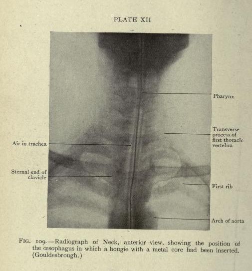
The image is a vintage radiographic view of the neck, taken from an anterior perspective. It appears to be a black and white photograph of an X-ray. The image shows the skeletal structure of the neck and upper chest, including the clavicles, ribs, and vertebrae. A bougie with a metal core is visible within the esophagus, appearing as a thin, bright line running down the center of the neck. Labels point to specific anatomical features, including the pharynx, transverse process of the first thoracic vertebra, first rib, arch of aorta, air in the trachea, and sternal end of clavicle. The image is labeled as 'FIG. 109 - Radiograph of Neck, anterior view, showing the position of the oesophagus in which a bougie with a metal core had been inserted.' and 'Plate XII'. The image has a vintage, slightly grainy quality, typical of older radiographic images.