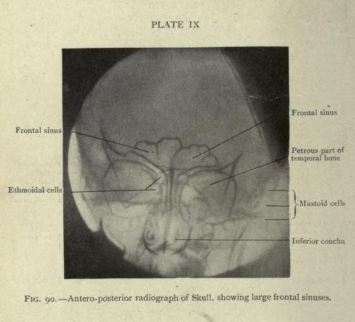
The image is a black and white radiograph of a skull, viewed from an antero-posterior perspective. It's labeled 'Plate IX' and 'Fig. 90 - Antero-posterior radiograph of Skull, showing large frontal sinuses'. The radiograph shows the internal structures of the skull, with several key areas labeled: * **Frontal Sinus:** Two large, dark, rounded areas are visible in the upper portion of the skull, representing the frontal sinuses. * **Ethmoidal Cells:** A complex network of smaller, darker areas is visible between and around the eyes, indicating the ethmoidal cells. * **Petrous Part of Temporal Bone:** A dense, bone-like structure is visible on either side of the skull, representing the petrous part of the temporal bone. * **Mastoid Cells:** A series of smaller, darker areas are visible behind the ears, indicating the mastoid cells. * **Inferior Concha:** A curved, bone-like structure is visible in the lower portion of the nasal cavity, representing the inferior concha. The overall image is a detailed anatomical representation of the skull's internal structures, as seen through radiography.