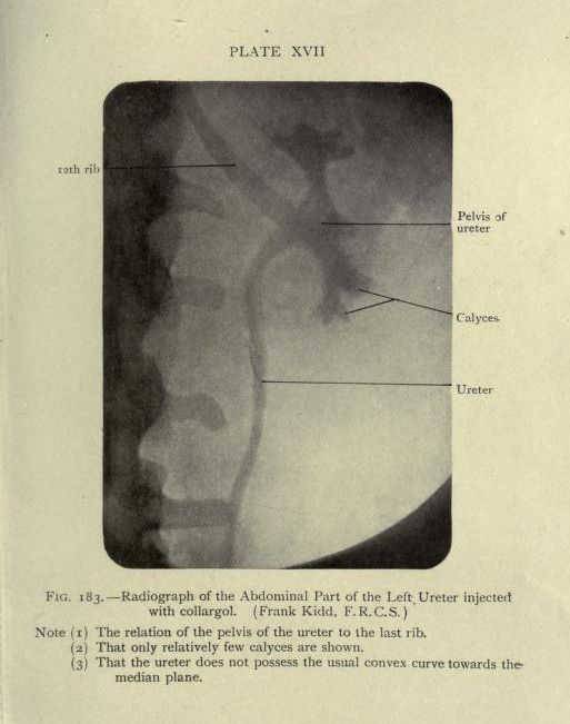
The image is a vintage medical radiograph titled 'Plate XVII' and labeled 'Fig. 183 – Radiograph of the Abdominal Part of the Left, Ureter injected with collargol.' It depicts a grayscale X-ray of a portion of the human anatomy, specifically the left ureter. The image shows a curved, tubular structure, the ureter, running vertically through the center. It's labeled with a line pointing to it. Above the ureter, there's a wider, cup-shaped structure labeled 'Pelvis of ureter'. Small, branching structures, labeled 'Calyces', are visible near the top of the image, connected to the pelvis of the ureter. A horizontal line indicates the '12th rib' at the top left of the image. The radiograph appears to be an older medical illustration, with a slightly grainy texture and a limited tonal range. There are notes below the image: (1) The relation of the pelvis of the ureter to the last rib. (2) That only relatively few calyces are shown. (3) That the ureter does not possess the usual convex curve towards the median plane.