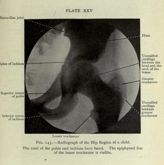
This is a vintage radiographic image of the hip region of a child. The image is a black and white X-ray, showing the bony structures of the pelvis and upper femur. The image is labeled with anatomical terms, pointing to specific features. Key features visible include: * **Pelvis:** The ilium, ischium, and pubis are clearly visible, forming the pelvic girdle. * **Femur:** The head and neck of the femur are visible, articulating with the pelvis. * **Trochanters:** The greater and lesser trochanters of the femur are prominent bony landmarks. * **Cartilage:** Areas of unossified cartilage are indicated between the head and neck of the femur, and between the greater trochanter and the femur. * **Rami:** The superior and inferior rami of the pubis and ischium are fused. * **Sacro-iliac joint:** The joint between the sacrum and ilium is visible. * **Epiphyseal line:** The epiphyseal line of the lesser trochanter is visible. The image is labeled with anatomical terms, pointing to specific features. The overall impression is a detailed anatomical study of a child's hip joint, likely for medical or educational purposes.