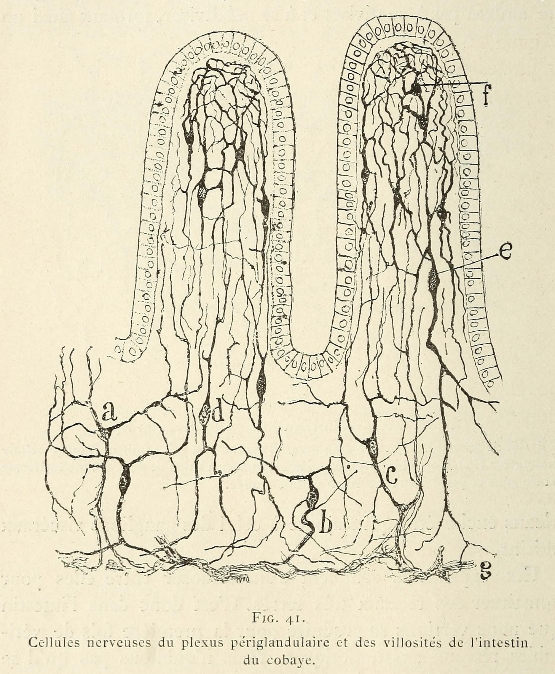
This is a vintage scientific illustration depicting nerve cells within the intestinal tissue of a guinea pig. The illustration is a detailed diagram of the periglandular plexus and villi of the intestine. It shows elongated, finger-like structures (villi) with a network of nerve cells and fibers surrounding them. The nerve cells are represented as small circles with branching extensions, forming a complex web. The illustration is done in black and white line art, with labels 'a', 'b', 'c', 'e', 'f', and 'g' pointing to different parts of the structure. The text below the illustration reads 'Cellules nerveuses du plexus périglandulaire et des villosités de l'intestin du cobaye', which translates to 'Nerve cells of the periglandular plexus and villi of the guinea pig intestine'.