radiographhipchildbonescartilageiliumischiumhead of femurfemursacro-iliac jointbody of pubisinferior ramus of ischiumradiographhipchildanatomyskeletonmedical illustrationvintageblack and white
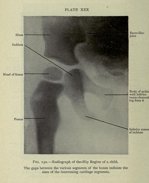
The image is a vintage black and white radiograph of the hip region of a child. It appears to be a medical illustration from a textbook or scientific publication, as indicated by the text at the bottom. The radiograph shows the skeletal structure of the hip, including the ilium, ischium, head of the femur, femur, sacro-iliac joint, body of pubis, and inferior ramus of the ischium. The bones are visible as lighter shades against a darker background. Labels with arrows point to specific anatomical features. The text below the image reads: 'FIG. 150. Radiograph of the Hip Region of a child. The gaps between the various segments of the bones indicate the sizes of the intervening cartilage segments.' The image is labeled 'PLATE XXX' at the top.
License: CC0