diagramcellnerve cellcell bodydendriteaxongray matterspinal cordfrogadultnervetissuebiological illustrationscientific diagramanatomyneurologymicroscopic viewcell structurebiological researchscientific studyillustrationdrawingsketchline artblack and whitevintagehistoricaltextlabelletterfigurediagramscientific illustrationbiological diagramanatomy diagramneurological diagrammicroscopic diagramcell diagramtissue diagramnerve diagramspinal cord diagramfrog diagramadult diagramnerve cell diagramcell body diagramdendrite diagramaxon diagramgray matter diagramscientific researchbiological studyanatomy studyneurology studymicroscopic studycell studytissue studynerve studyspinal cord studyfrog studyadult studynerve cell studycell body studydendrite studyaxon studygray matter studyfrogspinal cordnerve cellsanatomyneurologyscientific illustrationvintagehistoricalbiologydiagramillustrationdrawingsketchline artblack and whitetissuecellnervedendriteaxongray matter
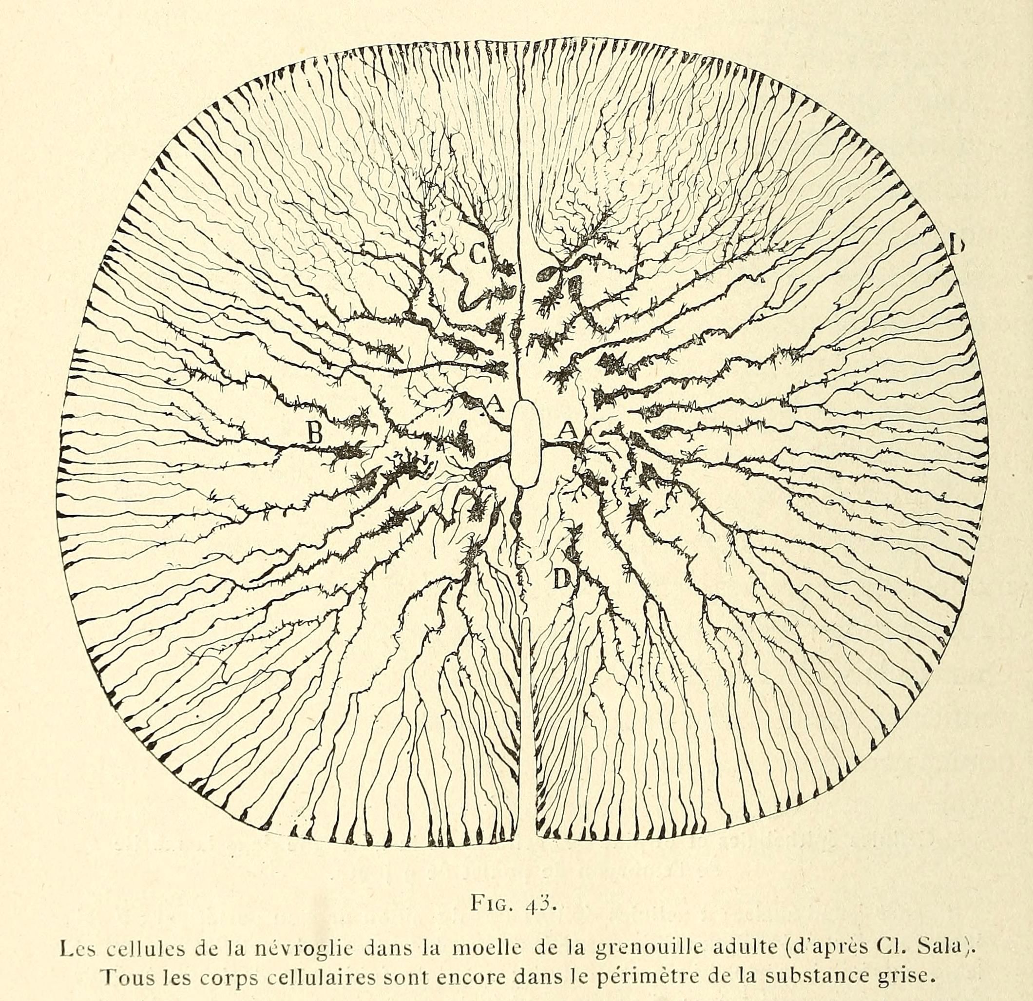
The image is a vintage black and white scientific illustration, likely a diagram of a frog's spinal cord. It depicts a circular cross-section of the spinal cord, with a central area labeled 'A' and radiating structures extending outwards. These structures resemble nerve cells with branching dendrites and axons. The illustration is highly detailed, showing the intricate network of cells and fibers within the spinal cord. Labels 'B', 'C', and 'D' are also present, likely indicating specific areas or structures within the diagram. The overall style is reminiscent of anatomical illustrations from the late 19th or early 20th century. Below the diagram, there is text in French, providing a caption or description of the illustration. The image is a valuable resource for studying the anatomy and structure of the frog's spinal cord.
License: CC0