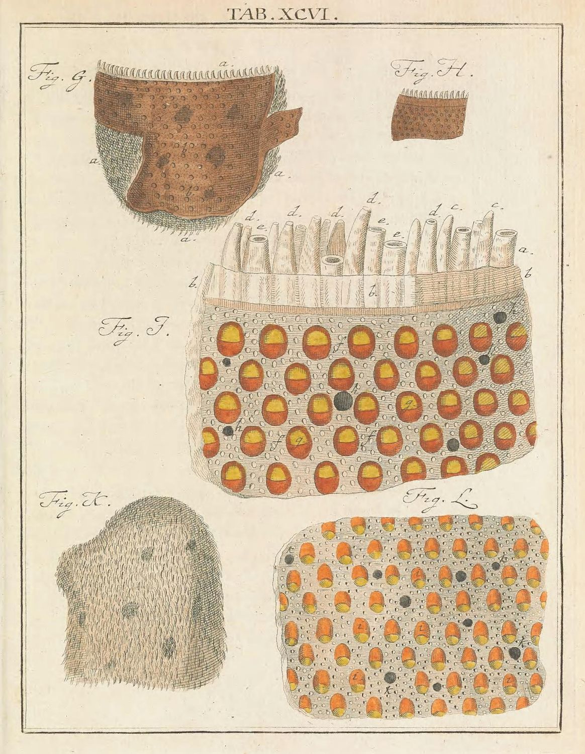
This is a vintage scientific illustration, likely from a medical or biological text. It depicts various stages or views of a parasitic mite infestation, specifically scabies. The illustration is composed of five separate figures labeled Fig. G, Fig. H, Fig. I, Fig. K, and Fig. L. * **Fig. G:** Shows a cross-section of a scalp covered in hair. The skin is marked with numerous small, dark dots representing the mites and their burrows. Labels 'a' and 'b' point to different layers of the skin. * **Fig. H:** A close-up, almost microscopic view of the skin, densely populated with the mites. It appears as a textured surface covered in tiny, dark organisms. * **Fig. I:** A larger view of a skin patch, also densely covered with the mites. The mites are more clearly visible as small, oval-shaped creatures. * **Fig. K:** A section of skin showing a more sparse distribution of mites, allowing for a clearer view of their shape and size. * **Fig. L:** A magnified view of the skin surface, showing the mites embedded within the skin's texture. The mites are depicted as small, oval-shaped creatures with visible legs. The illustration is rendered in a detailed, precise style, typical of scientific illustrations from the 18th or 19th century. The use of line work and shading creates a sense of depth and texture. The overall tone is informative and educational, aimed at providing a visual understanding of the scabies infestation.