diagramillustrationdrawingbotanical illustrationscientific illustrationmicroscopic viewplant structurecellular structurecross-sectionstemleafbundlefibercelltissuevascular bundlexylemphloembundle sheathcell wallcytoplasmnucleuscell shapecell arrangementplant anatomybotanyscientific studyresearchdocumentationhistorical illustrationvintage illustrationengravingetchingblack and whitemonochromedetailed drawingprecise illustrationlabeled diagramnumbered diagramlettered diagramscientific diagramplatefigurediagrammatic representationvisual aideducational materialtextlabelannotationborderframepagedocumentarchivecollectionhistorical recordscientific archiveresearch archivevintage documentantique documenthistorical illustrationscientific illustrationbotanical illustrationmicroscopic viewplant structurecellular structurecross-sectionstemleafbundlefibercelltissuevascular bundlexylemphloembundle sheathcell wallcytoplasmnucleuscell shapecell arrangementplant anatomybotanyscientific studyresearchdocumentationhistorical illustrationvintage illustrationengravingetchingblack and whitemonochromedetailed drawingprecise illustrationlabeled diagramnumbered diagramlettered diagramscientific diagramplatefigurediagrammatic representationvisual aideducational materialtextlabelannotationborderframepagedocumentarchivecollectionhistorical recordscientific archiveresearch archivevintage documentantique documentplant anatomybotanymicroscopic viewcellular structurevascular bundlexylemphloemstemleaftissuecellscientific illustrationvintage illustrationhistorical illustrationdiagramdrawingengravingetching
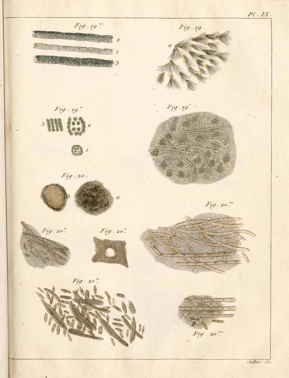
This is a vintage scientific illustration, likely from a botanical or biological text. The page is filled with detailed, black and white diagrams of plant structures at a microscopic level. There are multiple figures labeled with numbers and letters (e.g., Fig. 19, Fig. 20, a, b, 1, 2, 3). The illustrations depict cross-sections of stems and leaves, showing vascular bundles, cells, and tissues. The drawings are precise and detailed, with shading to indicate depth and texture. The style is typical of scientific illustrations from the 19th or early 20th century. The page has a slightly aged appearance, with a subtle texture to the paper. At the top right corner, it reads 'Pl. IX', indicating it's part of a larger plate or series. At the bottom right corner, it reads 'Sellier Sc.' which is likely the name of the illustrator or engraver.
License: CC0