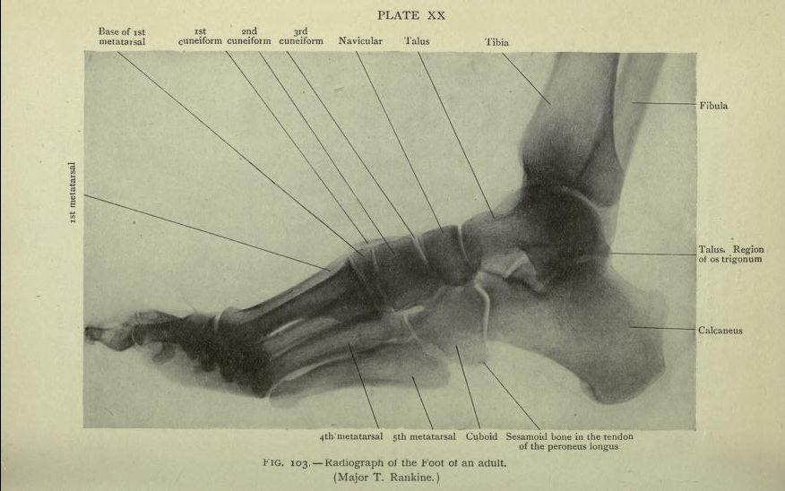
This is a black and white radiograph (X-ray) of an adult foot, labeled with anatomical terms. The image shows a side view of the foot and lower leg. The bones are clearly visible, appearing as lighter shades against the darker background. The tibia and fibula are visible in the upper portion of the image, forming the lower leg. Below these, the talus, calcaneus (heel bone), and navicular are prominent. The cuneiform bones (1st, 2nd, and 3rd) are also labeled, and the metatarsal bones are visible extending towards the toes. The 4th and 5th metatarsals are specifically labeled. A small sesamoid bone is noted within the tendon of the peroneus longus. Labels with lines point to the specific bones, providing anatomical identification. The image is titled 'FIG. 103 - Radiograph of the Foot of an adult' and is labeled 'PLATE XX' with the name 'Major T. Rankine' below.