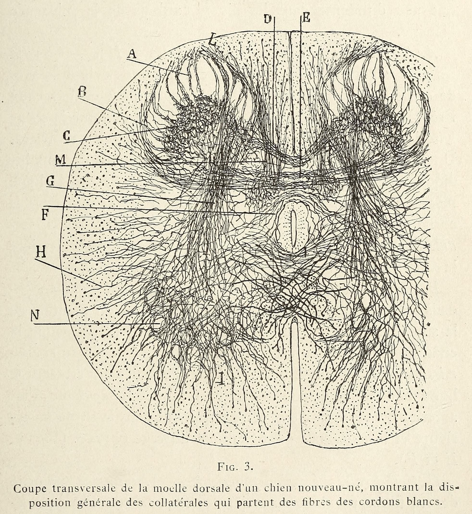
This is a vintage anatomical diagram depicting a transverse section of the dorsal spinal cord of a newborn puppy. The image is a black and white illustration with a dense network of fine lines representing nerve fibers and their collateral branches. The overall shape is roughly oval, representing the spinal cord's cross-section. The majority of the image is filled with these intricate lines, radiating outwards and crisscrossing each other. The lines are denser in the central region and become more sparse towards the periphery. This illustrates the distribution of nerve fibers and their connections within the spinal cord. Several labels with letters (A, B, C, D, E, F, G, H, M, N) are placed around the diagram, pointing to specific areas or structures within the spinal cord. These labels likely correspond to descriptions in the accompanying text. Below the diagram, there is text in French: 'Coupe transversale de la moelle dorsale d'un chien nouveau-né, montrant la disposition générale des collatérales qui partent des fibres des cordons blancs.' This translates to 'Transverse section of the dorsal spinal cord of a newborn puppy, showing the general arrangement of the collaterals originating from the fibers of the white matter.' The style of the illustration is typical of anatomical drawings from the late 19th or early 20th century, with a focus on detailed representation of structures and a somewhat stylized aesthetic.