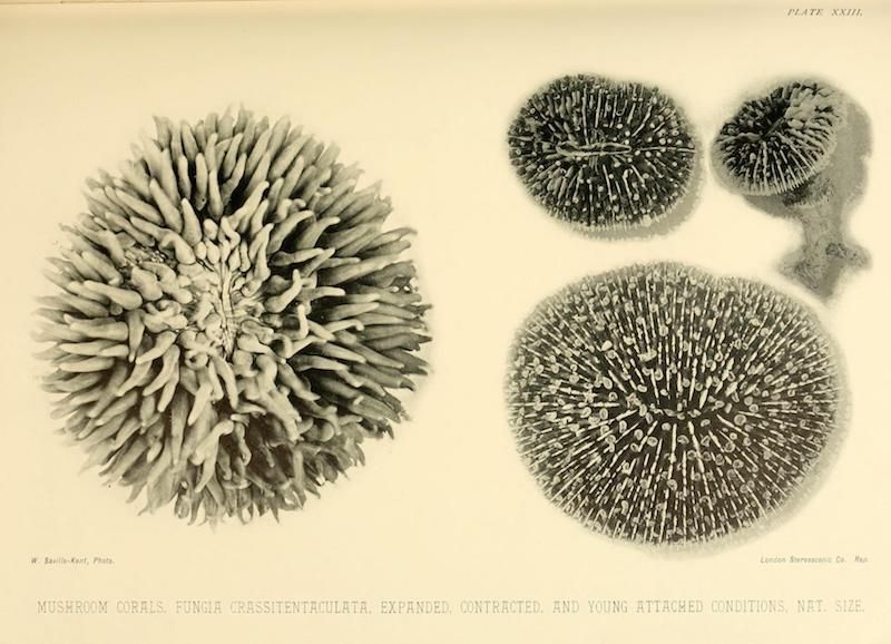
The image is a vintage scientific illustration depicting various stages of a mushroom coral, *Fungia crassitentaculata*. It's a plate from a larger publication, labeled 'Plate XXIII' at the top. The background is a creamy, aged paper color. There are four depictions of the coral in different states: 1. **Large Expanded Coral:** On the left, a large, detailed illustration shows the coral fully expanded. It's a rounded, dome-like shape with numerous, finger-like tentacles radiating outwards. The tentacles are densely packed and have a slightly textured appearance. The color is a muted gray-green. 2. **Contracted Coral:** Above the expanded coral, a smaller illustration shows the coral in a contracted state. It appears as a more compact, rounded shape with the tentacles drawn in closer. The color is similar to the expanded coral. 3. **Young Attached Coral:** To the right of the contracted coral, a very small illustration shows a young coral attached to a surface. It's a tiny, rounded shape with a few visible tentacles. 4. **Large Contracted Coral:** Below the expanded coral, a large illustration shows the coral in a contracted state. It appears as a more compact, rounded shape with the tentacles drawn in closer. The color is similar to the expanded coral. Below the illustrations, there's text identifying the subject as 'MUSHROOM CORALS. Fungia crassitentaculata. Expanded, Contracted, and Young Attached Condition. Nat. Size.' There are also publisher details in the bottom corners. The style is detailed and realistic, typical of scientific illustrations from the late 19th or early 20th century. The shading and texture are created using fine lines and stippling.