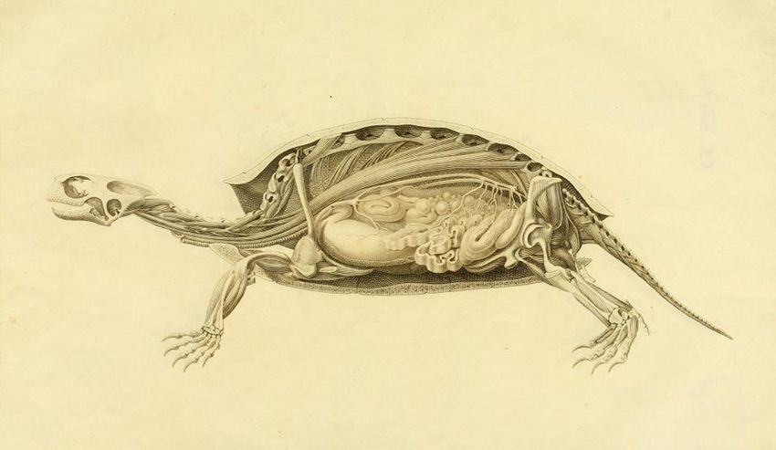
This is a detailed anatomical illustration of a turtle. The turtle is depicted in a side view, with a significant portion of its shell removed to reveal its internal organs and skeletal structure. The shell itself is outlined, showing the individual scutes (plates) that make up its carapace and plastron. Beneath the shell, a complex network of muscles, bones, and organs is visible. The skeletal structure, including the ribs, spine, and limb bones, is clearly defined. The internal organs, such as the lungs, heart, liver, and intestines, are rendered with a level of detail that suggests a scientific or educational purpose. The illustration is executed in a style reminiscent of vintage anatomical drawings, with fine lines and shading used to create depth and texture. The overall color palette is muted, with shades of brown and beige dominating the image. The background is a pale yellow, which helps to highlight the details of the turtle's anatomy. The image appears to be a historical or scientific illustration, likely intended for use in a textbook or research publication. It provides a detailed and accurate representation of the turtle's internal structure, making it a valuable resource for students and researchers alike.