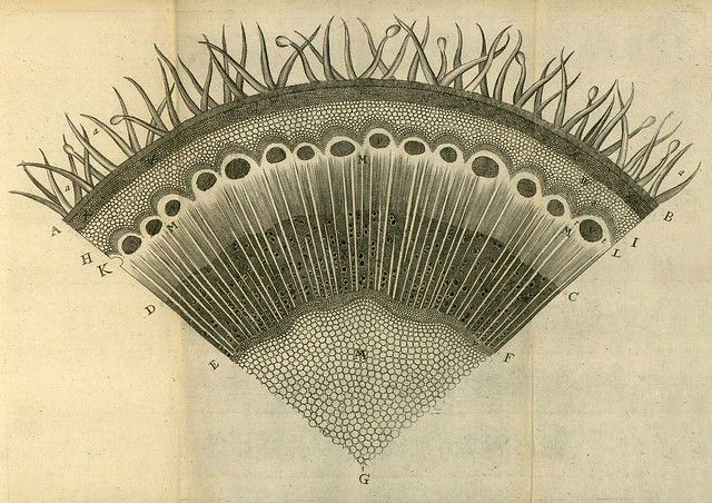
This is a detailed, vintage botanical illustration depicting a cross-section of a leaf. The illustration is rendered in a precise, engraved style, typical of scientific illustrations from the 18th or 19th century. The leaf is shown as a semi-circular segment, revealing its internal structure. The outermost layer is the epidermis, with a waxy cuticle on the upper surface. Below this, the mesophyll is visible, composed of tightly packed cells. Within the mesophyll, there are numerous vascular bundles, appearing as darker, elongated structures. These bundles contain xylem and phloem for transporting water and nutrients. Scattered throughout the mesophyll are stomata, small pores used for gas exchange, appearing as oval openings. The cells within the leaf are densely packed, with a stippled texture to indicate their cellular structure. Numerous labels, indicated by letters (A, B, C, D, E, F, G, H, I, K, L, M, N), point to different parts of the leaf's anatomy. The background is a faded, aged paper color, adding to the vintage aesthetic. The overall impression is one of scientific accuracy and detailed observation.