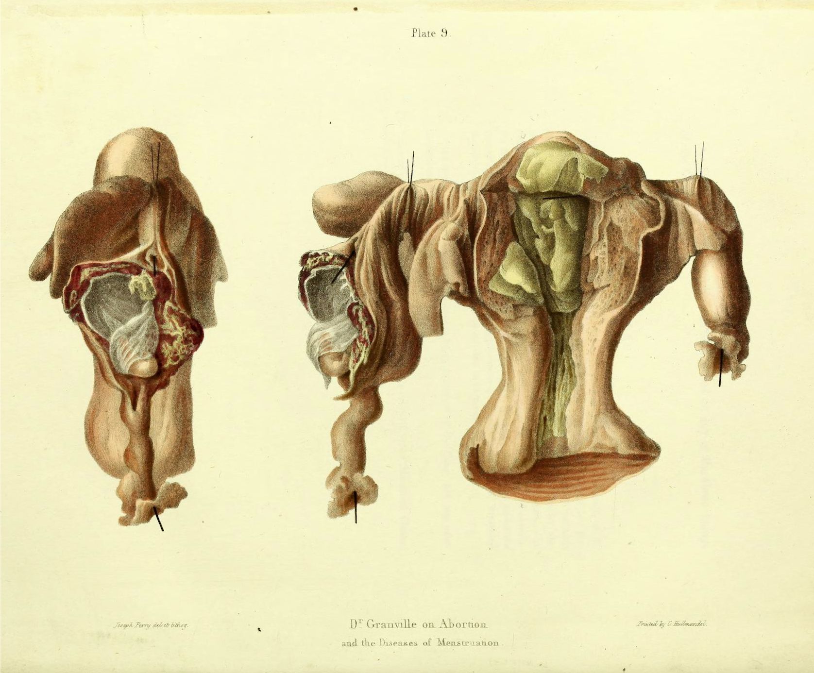
This is a vintage anatomical illustration, likely from a medical textbook, depicting the female reproductive system. It features two views of a dissected uterus, fallopian tubes, and ovaries. The illustration is rendered in a detailed, realistic style with watercolor-like coloring. The uterus is the central focus, shown in a pale pinkish-beige tone. The fallopian tubes extend from the uterus, and the ovaries are visible on either side. The internal structure of the uterus is exposed, revealing a complex network of blood vessels and tissue. Arrows point to specific anatomical features, likely for educational purposes. The illustration is labeled with the title "D. Granville on Abortion and the Diseases of Menstruation" and the plate number "Plate 9". The artist's name, "J. Harris", is also visible. The background is a pale yellow, and the overall impression is one of scientific accuracy and historical significance.