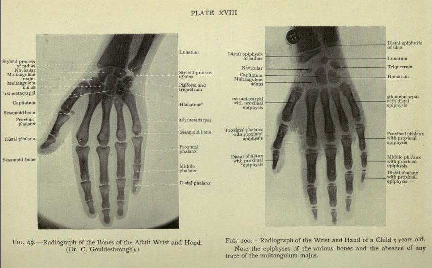
The image is a plate from a medical textbook, showing two radiographs of the hand and wrist. The plate is titled 'PLATE XVIII'. **Left Side (Fig. 99):** This is a radiograph of the bones of an adult wrist and hand. The bones are clearly visible as white shapes against a dark background. Labels point to specific bones, including the 'styloid process of radius', 'navicular', 'multangulum majus', 'capitatum', 'sesamoid bone', 'proximal phalanx', 'digital phalanx', '5th metacarpal', and others. The bones are depicted in a realistic anatomical arrangement. **Right Side (Fig. 100):** This is a radiograph of the wrist and hand of a 5-year-old child. The bones are similar to the adult hand, but with noticeable differences. The labels point to the same bones as the adult hand, but with the addition of 'epiphysis' labels on some bones, indicating the growth plates present in a child's developing skeleton. The text notes the presence of epiphyses and the absence of the 'multangulum majus'. **Overall:** The image is a comparative anatomical illustration, demonstrating the differences between the skeletal structure of an adult and a child's hand and wrist. The radiographs are detailed and labeled, making it a useful educational tool for medical students or anyone interested in anatomy. The background is a light beige color, and the image has a vintage, textbook-like appearance.