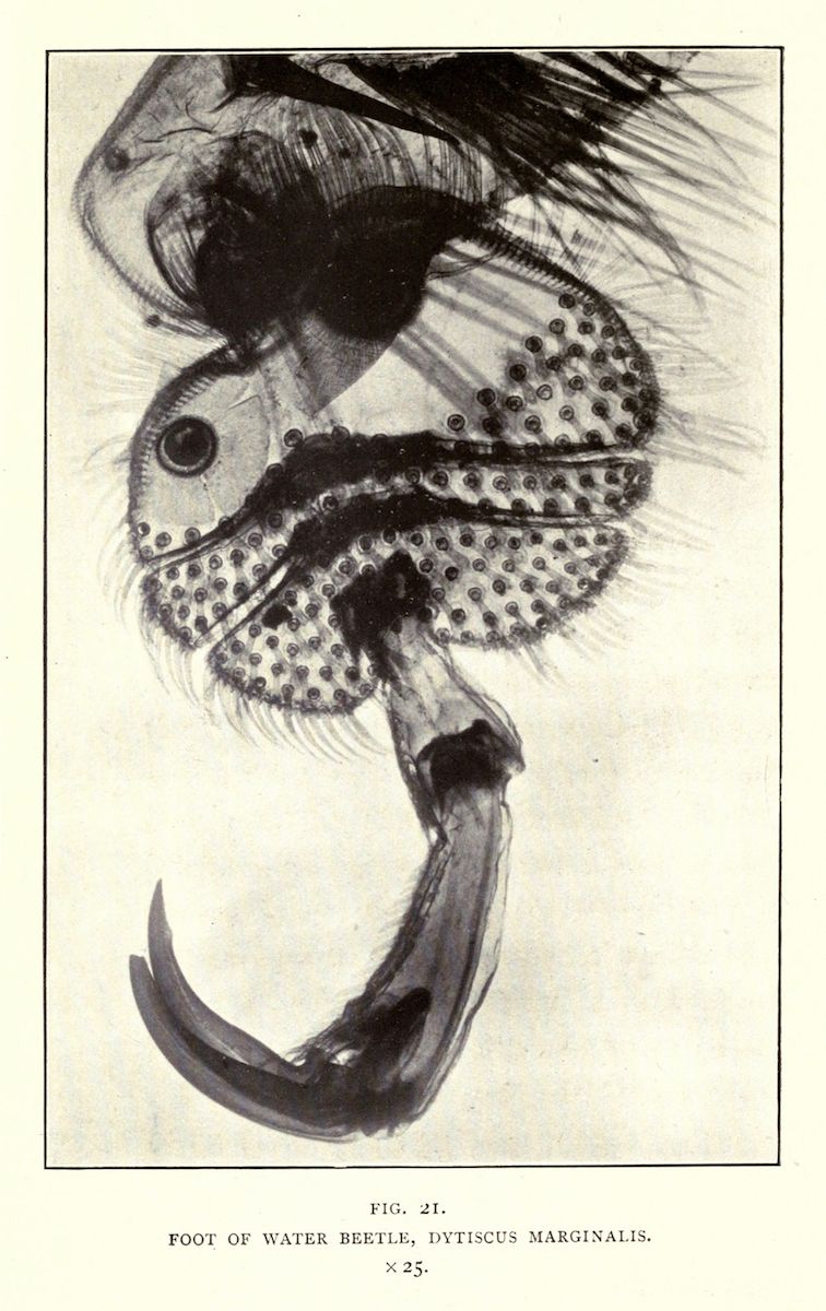
The image is a detailed black and white illustration of the foot of a water beetle, specifically identified as *Dytiscus marginalis*. The foot is highly magnified (indicated by 'x 25' at the bottom) and appears as a complex structure with numerous small, rounded protrusions covering its surface. These protrusions resemble tiny suction cups or bristles. The foot is attached to a slender, curved leg. At the top of the leg, there's a small, dark eye-like structure. The illustration is a scientific drawing, likely from a biological or entomological publication, emphasizing the intricate details of the beetle's foot. The background is plain white, highlighting the subject. The text at the bottom reads 'FIG. 21. FOOT OF WATER BEETLE, DYTISCUS MARGINALIS.'