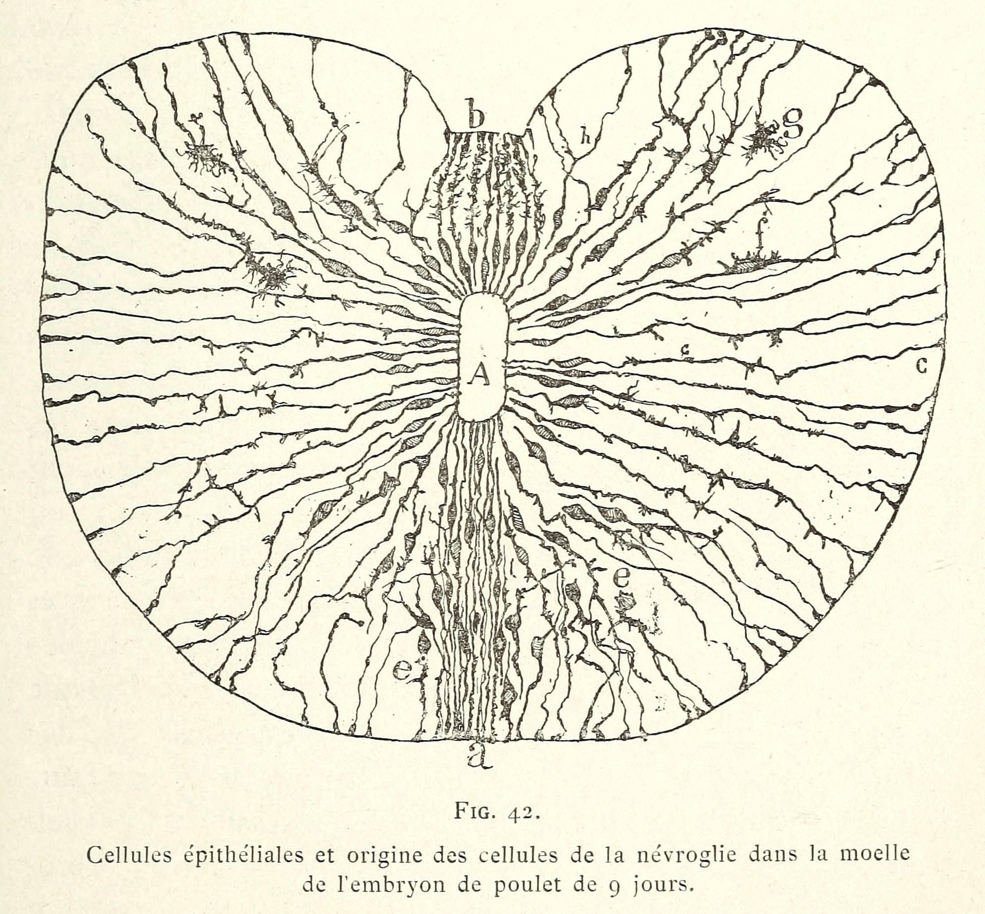
The image is a vintage scientific illustration, likely from a biology or embryology textbook. It's a diagram of a 9-day-old chicken embryo's neural crest cells and their origin within the marrow. The central focus is a roughly oval shape, representing a cross-section of the embryo's tissue. Within this shape, a complex network of lines radiates outwards from a central point labeled 'a'. These lines represent the migration pathways of the neural crest cells. The lines are densely packed near 'a' and become more sparse as they extend towards the edges of the oval. Several other labels are present: 'b', 'c', and 'e' are indicated with arrows pointing to specific areas within the diagram. The lines are drawn in a detailed, almost woodcut style, with a lot of fine detail. Below the diagram, there is text in French: 'Cellules épithéliales et origine des cellules de la névroglie dans la moelle de l'embryon de poulet de 9 jours.' This translates to 'Epithelial cells and origin of neuroglia cells in the marrow of the 9-day-old chicken embryo.' The overall aesthetic is that of a classic scientific illustration, with a focus on clarity and detail.