radiographwristradiusulnaboneshandanatomymedical imageradiographx-raywristanatomybonesradiusulnacarpalsmetacarpalssesamoid bonesmedical illustrationvintageblack and white
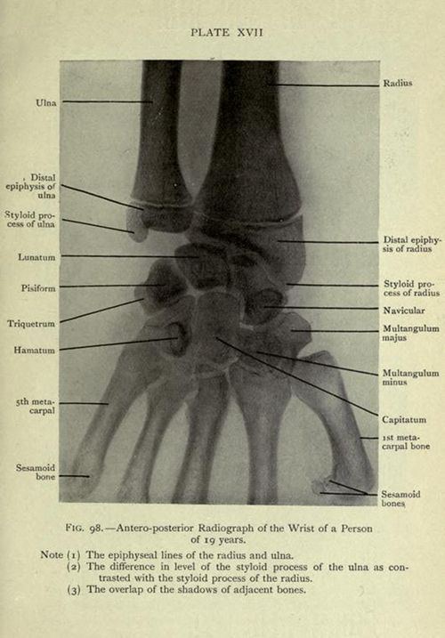
This is a vintage black and white radiograph (X-ray) of a human wrist, labeled with anatomical terms. The image shows the bones of the forearm (radius and ulna) extending into the wrist and hand. The radius and ulna are clearly visible, with labels pointing to their distal epiphyses and styloid processes. The wrist bones (carpals) are also labeled, including the lunate, pisiform, triquetrum, hamatum, capitate, and various metacarpal bones. Sesamoid bones are also indicated. The image is a medical illustration used for anatomical study, with notes below the image highlighting specific features like the epiphyseal lines and the overlap of shadows between bones.
License: CC0