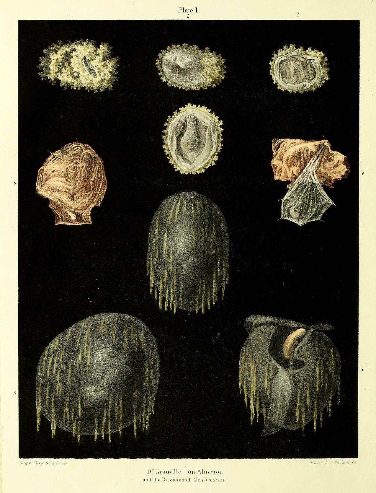
This is a vintage medical illustration, likely from a textbook or anatomical study. It's titled 'Plate 1' and labeled 'D. Granville on Abortion and the Diseases of Menstruation' at the bottom. The background is a deep, inky black. Nine detailed diagrams are arranged in a 3x3 grid. Each diagram depicts a cross-section of what appears to be a uterus, likely at different stages of pregnancy or related to conditions affecting the reproductive system. The illustrations are rendered in shades of brown, beige, and white, with a delicate, almost watercolor-like quality. Each uterine depiction is unique, showing varying degrees of development or abnormalities. Some show a smooth, rounded shape, while others exhibit irregularities, growths, or internal structures. Many have a fringe-like texture around the edges, possibly representing tissue or membranes. The illustrations are highly detailed, with careful attention paid to the textures and shapes of the internal organs. The overall impression is one of scientific accuracy and historical significance. The illustrations are likely intended to educate medical professionals or students about the anatomy and pathology of the female reproductive system.