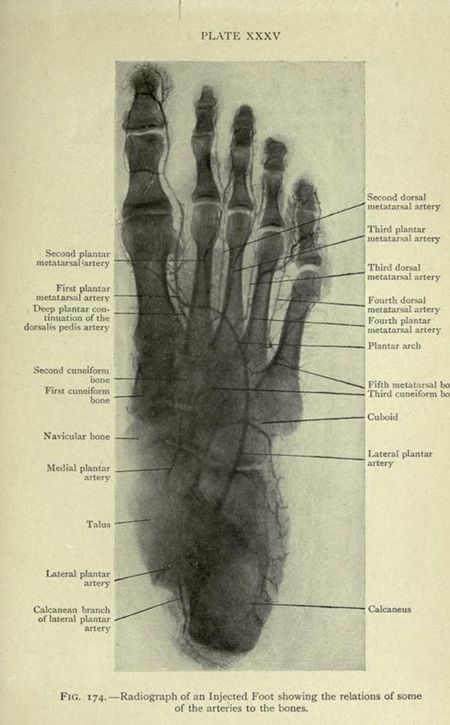
This is a vintage radiographic image of an injected human foot, labeled with anatomical terms. The image is a black and white plate labeled 'PLATE XXXV' at the top. The foot is depicted in a dorsal view, showing the bones and a network of arteries that have been injected to make them visible. The bones of the foot are clearly visible, including the talus, calcaneus, navicular bone, cuneiform bones (first and second), cuboid, and metatarsal bones. The toes are also visible, though less detailed. A complex network of arteries is visible throughout the foot, branching and winding around the bones. These arteries are labeled with anatomical terms such as 'dorsalis pedis artery', 'lateral plantar artery', 'medial plantar artery', 'first plantar metatarsal artery', 'second plantar metatarsal artery', 'third plantar metatarsal artery', 'fourth plantar metatarsal artery', 'second dorsal metatarsal artery', 'third dorsal metatarsal artery', 'fourth dorsal metatarsal artery', 'calcanean branch of lateral plantar artery', and others. Below the image, there is a caption that reads 'FIG. 174. Radiograph of an Injected Foot showing the relations of some of the arteries to the bones'. The overall impression is that of a detailed anatomical illustration used for medical or educational purposes.