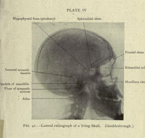
This is a vintage black and white radiograph (X-ray) of a human skull, presented in a side view. The image is labeled with anatomical features, indicated by lines pointing to specific areas of the skull. Here's a breakdown of the labeled features: * **Hypophyseal fossa (pituitary):** Located within the skull, near the center. * **Sphenoidal sinus:** Situated behind the nasal cavity. * **Frontal sinus:** Above the nasal cavity, in the forehead region. * **Ethmoidal cells:** Located between the eyes, within the ethmoid bone. * **Maxillary sinus:** Below the eyes, in the cheekbone area. * **Internal acoustic meatus:** A canal in the temporal bone. * **Condyle of mandible:** The rounded projection of the lower jaw. * **Floor of tympanic antrum:** Part of the middle ear. * **Atlas:** The first cervical vertebra, visible in the lower part of the image. The image has a grainy, vintage quality, typical of older radiographic images. The skull is depicted in profile, showing the contours of the cranial bones and the internal structures as they appear on the radiograph. The image is labeled 'PLATE IV' and 'FIG. 41. - Lateral radiograph of a living Skull.'