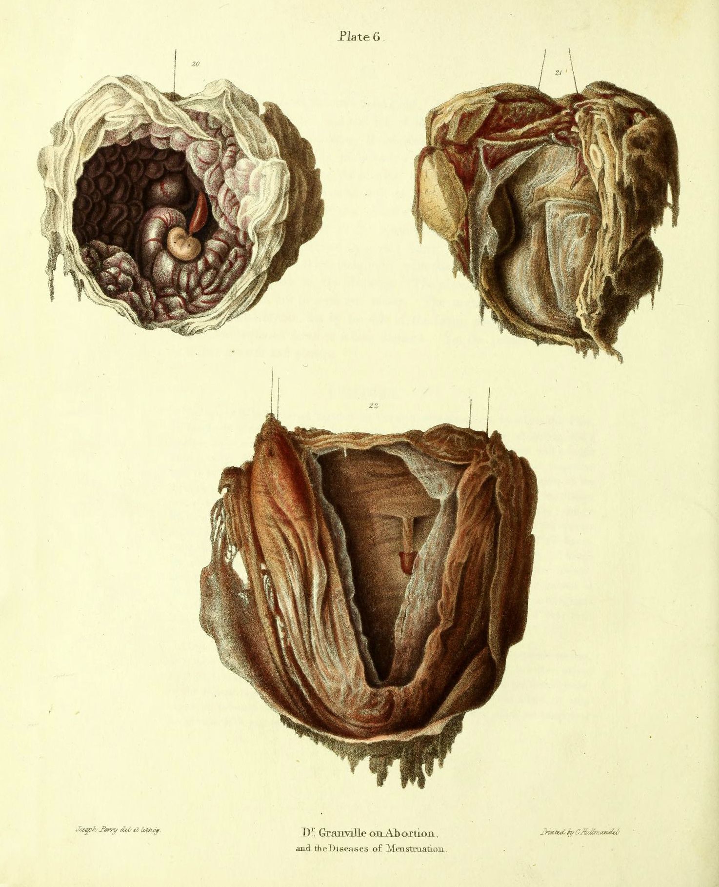
This is a vintage anatomical illustration, likely from a medical textbook, depicting three different views of a uterus with a fetus inside. The background is a pale yellow. **Top Left:** A cross-section of a uterus is shown, revealing a fetus within. The fetus is small and appears to be in a curled position. The uterine lining is visible, with a network of blood vessels. The placenta is also visible, attached to the uterine wall. **Top Right:** A view of the uterus from the outside, showing the exterior surface. The texture is rough and irregular, with visible blood vessels and folds of tissue. **Bottom:** Another view of the uterus, cut open to reveal the interior. The fetus is visible, surrounded by the placenta and amniotic fluid. The uterine wall is thick and muscular, with a network of blood vessels. Each illustration is labeled with a number (20, 21, 22) and there is text at the bottom of the image that reads “Dr Granville on Abortion and the Diseases of Menstruation”. The illustration is signed by “J. Perry del et lith” and “Printed by C. Hullmandel”. The overall style is detailed and realistic, typical of anatomical illustrations from the 19th century. The colors are muted, with shades of pink, red, and brown.