radiographhipchildsacroiliac jointiliumunossified cartilagepubisischiumhead of femurgreater trochanterlesser trochanterradiographhipchildanatomyskeletonbonescartilagemedical imagingpediatrics
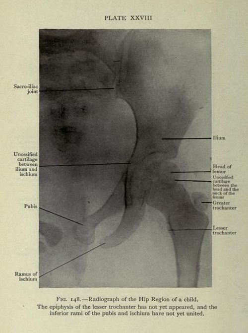
The image is a vintage black and white radiograph of the hip region of a child. It's labeled with anatomical terms pointing to various bones and cartilaginous areas. The radiograph shows the sacroiliac joint, ilium, pubis, ischium, and the head of the femur. Areas of unossified cartilage are indicated between the ilium and ischium, and between the head and neck of the femur. The greater and lesser trochanters are also labeled. A caption at the bottom states that the epiphysis of the lesser trochanter has not yet appeared, and the inferior rami of the pubis and ischium have not yet united. The image is labeled 'Plate XXVIII' and 'Fig. 148'.
License: CC0