bonekneeradiographfemurpatellatibiafibulasesamoid bonegastrocnemiusmedial condyle of femurkneeradiographx-rayanatomybonesfemurpatellatibiafibulamedical imagingorthopedics
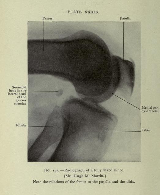
This is a vintage black and white radiograph (X-ray) of a fully flexed knee. The image shows the bones of the knee joint, including the femur (thigh bone), patella (kneecap), tibia (shin bone), and fibula. The bones are clearly visible due to the contrast of the radiograph. Labels point to specific anatomical features: the femur, patella, tibia, fibula, a sesamoid bone in the head of the gastrocnemius muscle, and the medial condyle of the femur. The image is labeled as 'Plate XXXIX' and 'Fig. 185 - Radiograph of a fully flexed Knee' with a note indicating the relation of the femur to the patella and tibia.
License: CC0